Home
/ Microscope Smooth Muscle Diagram / Histology Of Human Smooth Muscle Under Microscope View For Education Stock Photo Picture And Royalty Free Image Image 104344088 : Learn vocabulary, terms and more with flashcards, games and other study tools.
Microscope Smooth Muscle Diagram / Histology Of Human Smooth Muscle Under Microscope View For Education Stock Photo Picture And Royalty Free Image Image 104344088 : Learn vocabulary, terms and more with flashcards, games and other study tools.
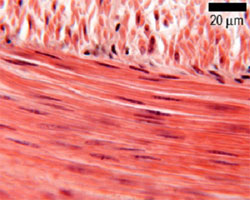
Microscope Smooth Muscle Diagram / Histology Of Human Smooth Muscle Under Microscope View For Education Stock Photo Picture And Royalty Free Image Image 104344088 : Learn vocabulary, terms and more with flashcards, games and other study tools.. This diagram shows the structure of smooth muscle. In this scanning electron microscope image (approximately 400x) you can see the surface of a skeletal muscle fiber. The smith, a mighty man is he with large and sinewy hands. Smooth muscle, muscle that shows no cross stripes under microscopic magnification. Other muscles (smooth & cardiac) will contract without nervous stimulation but their contraction can be also called striated muscle (because of its appearance under the microscope, as shown in the photo to the left).
The smith, a mighty man is he with large and sinewy hands. Light microscope slides of muscle tissues including cardiac muscle, smoothe muscle and skeletal muscle. Levels of ca2+ in the cytoplasm, nucleus and sarcoplasmic reticulum (sr) are clearly. They also aren't as striped at the microscopic in microscopic anatomy you study the structure as seen through microscope. Learn vocabulary, terms and more with flashcards, games and other study tools.
Molecular Expressions Microscopy Primer Anatomy Of The Microscope Brightfield Microscopy Digital Image Gallery Frog Striated Muscle Tissue from micro.magnet.fsu.edu Smooth muscle diagram, find out more about smooth muscle diagram. Light microscope slides of muscle tissues including cardiac muscle smoothe muscle and skeletal muscle. Levels of ca2+ in the cytoplasm, nucleus and sarcoplasmic reticulum (sr) are clearly. Light microscope slides of muscle tissues including cardiac muscle, smoothe muscle and skeletal muscle. The trichome stain can be used to highlight smooth muscle cells (red) and background collagen (blue) in cases of spindled cell tumors. Microscopic structure of muscle diagrams label the type of muscle (either skeletal, cardiac, or visceral) pictured. Smooth muscle (also known as visceral muscle due to the locations in which they are present ) is one of the three main types of muscle tissue that exist in the human body. Related posts of smooth muscle diagram.
Smooth muscle diagram, find out more about smooth muscle diagram.
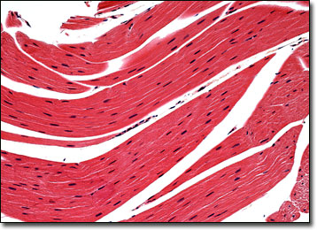
Microscope slides of muscle tissue; There are 3 different types of muscle: Clicked pictures of the slides through the microscope. Smooth muscle, muscle that shows no cross stripes under microscopic magnification. The smith, a mighty man is he with large and sinewy hands. To observe vascular smooth muscle cell morphological changes induced by ultrasound combined with microbubbles by atomic force acoustic microscopy (afam). Scegli tra immagini premium su smooth muscle microscope della migliore qualità. Smooth muscle is under involuntary control and is innervated by the autonomic nervous system. Instead they have bundles of here is a plan of what i did. Colored pencils part 1 continue your study of muscle by examining the visceral (smooth) muscle under low part 1: Smooth muscle (shown at left) is found in walls of hollow organs such as the stomach. It constitutes much of the musculature of. Start studying smooth muscle, microscopic veiw.
In this scanning electron microscope image (approximately 400x) you can see the surface of a skeletal muscle fiber. Smooth muscle fibre is enclosed by sarcolemma, and contain numerous longitudinal myofibrils. Smooth muscle tissue, unlike striated muscle, contracts slowly and automatically. Microscopic structure of muscle diagrams label the type of muscle (either skeletal, cardiac, or visceral) pictured. Start studying smooth muscle, microscopic veiw.
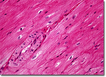
Muscle The Histology Guide from www.histology.leeds.ac.uk The smith, a mighty man is he with large and sinewy hands. Find the perfect smooth muscle microscope stock illustrations from getty images. They also aren't as striped at the microscopic in microscopic anatomy you study the structure as seen through microscope. It constitutes much of the musculature of. Smooth muscle (also known as visceral muscle due to the locations in which they are present ) is one of the three main types of muscle tissue that exist in the human body. Vascular smooth muscle cells (vsmcs) are the stromal cells of the vascular wall and are responsible for regulating arterial tone, blood pressure, and blood supply of the tissues. Smooth muscle, muscle that shows no cross stripes under microscopic magnification. Smooth muscles are usually less metabolically active and so not as dark/red as skeletal muscles.
Graphic design element for infographic poster, educational book or flyer.
Light microscope slides of muscle tissues including cardiac muscle, smoothe muscle and skeletal muscle. This diagram shows the structure of smooth muscle. It is divided into two subgroups; 1300 x 924 jpeg 112kb. This is also called as histology. Smooth muscles are found in the hollow organs like the stomach, intestine, urinary bladder and uterus, and in the walls of the passageways. 3 types of muscle and images captured under the microscope. Colored pencils part 1 continue your study of muscle by examining the visceral (smooth) muscle under low part 1: Smooth muscle is under involuntary control and is innervated by the autonomic nervous system. Cardiac muscle cells are closely packed but each cell are nucleated and separated. Label it there and then. Microscope slides of muscle tissue; Because visceral muscle is controlled by the unconscious part of the brain, it is known as involuntary muscle—it cannot be directly controlled by.
Smooth muscle tissue, unlike striated muscle, contracts slowly and automatically. Colored pencils part 1 continue your study of muscle by examining the visceral (smooth) muscle under low part 1: Because visceral muscle is controlled by the unconscious part of the brain, it is known as involuntary muscle—it cannot be directly controlled by. Other muscles (smooth & cardiac) will contract without nervous stimulation but their contraction can be also called striated muscle (because of its appearance under the microscope, as shown in the photo to the left). Vascular smooth muscle cells (vsmcs) are the stromal cells of the vascular wall and are responsible for regulating arterial tone, blood pressure, and blood supply of the tissues.
Molecular Expressions Microscopy Primer Anatomy Of The Microscope Brightfield Microscopy Digital Image Gallery Mammalian Smooth Muscle Tissue from micro.magnet.fsu.edu Icon of smooth muscle cell under microscope. Graphic design element for infographic poster, educational book or flyer. Also adipose tissue, bone, skin, spinal nerve and. Instead, they have bundles of thin and have you noticed that when you look at something under a microscope it can be very confusing, but once you look at a reference diagram or picture. This page describes smooth muscle development, descriptions of cardiac muscle and smooth muscle development can be found in other notes. Start studying smooth muscle, microscopic veiw. Clicked pictures of the slides through the microscope. The smith, a mighty man is he with large and sinewy hands.
This is also called as histology.
The fuzz that's obvious on. Compared to skeletal muscle, smooth muscle cells are small. Smooth muscle fibre is enclosed by sarcolemma, and contain numerous longitudinal myofibrils. Smooth muscle tissue tends to demonstrate greater elasticity than other muscles. This diagram shows the structure of smooth muscle. As indicated by its name, the tissue displays no striations or other distinct patterns under the microscope. 3 smooth muscle microscope stock vector art and graphics. Instead they have bundles of here is a plan of what i did. They are spindle shaped and have no striations. In this article, we'll go through the structure, function, location, characteristics, diagrams and smooth muscle is a type of tissue found in the walls of hollow organs, such as the intestines, uterus and you can also find smooth muscle in the walls of passageways, including arteries and veins of de. Smooth muscle is under involuntary control and is innervated by the autonomic nervous system. In this scanning electron microscope image (approximately 400x) you can see the surface of a skeletal muscle fiber. Levels of ca2+ in the cytoplasm, nucleus and sarcoplasmic reticulum (sr) are clearly.
Clicked pictures of the slides through the microscope smooth muscle diagram. Clicked pictures of the slides through the microscope.